Mandibular Canal Definition
It is a channel that extends from the mandibular foramen located on the median surface of the ramus, the posterior section of the mandible which is somewhat vertical in appearance.
Mandibular Canal Anatomy
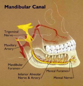
It is present inside the mandible that consists of:
- Inferior alveolar vein
- Inferior alveolar nerve
- Inferior alveolar artery
Mandibular Canal Contents
Apart from veins and arteries, it also consists of mandibular blood vessels as well as a part of the mandibular offshoot of the trigeminal nerve.
Mandibular canal Location
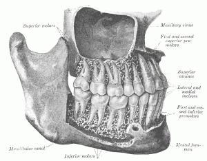
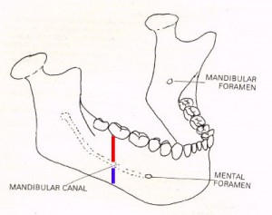
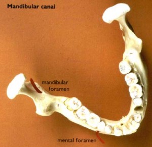
This internal canal runs transversely through the middle of the mandible and comes from posterior to anterior. It runs downward in an oblique manner and then forward in the ramus. It then moves forward horizontally inside the body, where it is located under the alveoli. It communicates with the alveoli through small apertures (openings). It runs parallel to the mandibular foramen and the mental foramen. It is the mandibular foramen that provides entry to the canal located in a posterior position to the ramus. Anteriorly, it provides an exit for the vessels and mental nerve from the canal.
Once the Mandibular canal reaches the incisor teeth, it reverts and communicates with the mental foramen. In this way, it emits a small canal called the mandibular incisive canal that runs to the cavities consisting of the incisor teeth.
Mandibular canal Function
This special channel carries branches of the inferior alveolar nerve, vessels, and artery that communicate with the dental alveoli through tiny passages.
Mandibular canal Names
The canal is known by other names like:
- Canalis mandibulae
- Canal mandibulaire
In Latin, it is also known as “Canalis mandibulae”. It is referred to as “Canal mandibulaire” in French.
Mandibular canal Etymology
The word Mandibular” comes from the term “mandible” which has been derived from the Latin verb “mandere” that means “to chew”.
Mandibular canal Variations
In 50% radiographs, the canal is found to be somewhat closely located to the vertices of the second molar. It is detected to be away from the root apices in 40% radiographs. In only 10%, the root vertices seem to get through the canal.
Mandibular canal Pathology
The canal houses vascular and neural tissue. Naturally, it is subject to pathology-related with the tissues. The channel exhibits neural malignancies as well as neural tumors, such as
- Schwannomas
- Neurofibromas
- Neuromas
Additionally, the canal is also found to be related to vascular tumors like
- A-V malformations
- Hemangiomas
- Malignancies, such as lymphomas
These types of pathology are often detected by the change in size, shape or location or course of the mandibular canal. When neural tumors are present, the mandibular canal suffers from an abnormal widening at the site of the tumor. The pathology of the canal is often diagnosed by an alteration in course, size or shape of the canal. Knowing these variations can help plan diagnostic and treatment procedures in the mandible and lead to fewer complications during treatment.
Mandibular Canal Pictures
Here are some useful Mandibular Canal photos for you. Take a look at these Mandibular Canal images to know how the channel looks like.

Picture 3 – Mandibular Canal Image

Picture 4 – Mandibular Canal Photo


No comments yet.