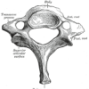
What is Vertebra prominens? It is the name given to the 7th cervical vertebra in the human body. Vertebra prominens Etymology The word “vertebra” is derived from the old Latin word “verto” meaning a “Joint”. It can also refer to something that is to be turned. In A.D. 30, this word was used by Celsus […]
Last updated on November 14th, 2018 at 8:14 pm
What is Frontalis? It is a thin, quadrilateral muscle that is attached closely to superficial fascia. It has got no bony attachments. It is sometimes considered to be a part of the occipitofrontalis muscle. (more…)
Last updated on August 21st, 2018 at 6:11 pm
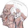
Levator Anguli Oris Definition It is a muscle responsible for elevating the angle of the mouth. It is connected to the nerve known as the buccal branch. This facial muscle receives fresh oxygenated blood from the facial artery network. Levator Anguli Oris Location This muscle arises from the Canine Fossa, just below the infraorbital foramen. […]
Last updated on February 2nd, 2018 at 7:29 am
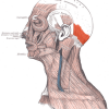
What is Occipitalis? The Occipitalis muscle is a thin, quadrilateral muscle that arises by the tendinous fibers from lateral two-thirds of occipital bone’s superior nuchal line as well as from the temporal bone’s mastoid section. Derived from Latin language, Occipitalis muscle is pronounced in Latin as Venter occipitalis musculi occipitofrontalis. Being one the skull covering […]
Last updated on August 23rd, 2018 at 1:43 pm

What is Suprasternal Notch? It is defined as a large, prominent dip on the apex of the sternum in the middle of articulation along with two clavicles. It is a significant division of the human anatomy. In adults, the notch is mainly present due to an aortic arch aneurysm. In a child, it is present […]
Last updated on July 24th, 2018 at 6:32 am
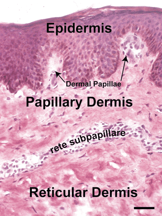
Papillary dermis Definition It is defined as the topmost layer of the dermis that entwines with the rete epidermis ridges. It is made up of fine collagen fibers that are arranged in a loose fashion. (more…)
Last updated on February 2nd, 2018 at 7:23 am
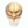
Preoccipital notch Definition It is a notch, or indentation, in the ventrolateral edge of the temporal lobe of either half of the cerebrum (Cerebral hemisphere). Preoccipital notch Synonyms It is known by a variety of names, like: Incisura praeoccipitalis Incisura preoccipitalis Incisura parieto-occipitalis Occipital notch Preoccipital incisure Preoccipital incisura Preoccipital notch Appearance It is approximately […]
Last updated on February 1st, 2018 at 12:55 pm
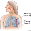
Visceral pleura Definition It is a thin serous membrane tissue layer that sticks to the lung surface. It is the innermost of the two pleural membrane layers investing the lungs. It is also known by the name Pulmonary pleura. In Latin, this structure is referred to as Pleura visceralis and Pleura pulmonalis. Visceral pleura Anatomy […]
Last updated on July 12th, 2018 at 7:54 am
Costal pleura Definition It is defined as the most extensive and strong section of the parietal pleural membrane. It is that part of the parietal pleura that lines the Intercostales and the inner rib surfaces. Costal pleura Location It lies adjacent to the intercostals muscles, costal cartilage, and the inner rib surface. Costal pleura Function […]
Last updated on February 1st, 2018 at 12:53 pm
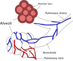
Terminal bronchiole Definition It is the terminal section of the nonrespiratory conducting airway. This bronchiole gets subdivided into respiratory bronchioles. The Terminal bronchioles are referred to as the last of the purely conductive airway generations in the human lungs. Terminal bronchiole Location It is located at the terminus of the conducting zone. It has a […]
Last updated on September 2nd, 2018 at 8:30 am


