What is Renal fascia?
It refers to a protective covering around each kidney. It is also known by the names “Gerota’s fascia” and “Gerota’s capsule.”
In Latin, it is referred to as “Fascia renalis.”
Renal fascia Anatomy
It is made up of connective tissue, a type of strong fibrous tissue that can be found all through the human body. The structure is surrounded by two layers of adipose tissue, a layer below and one above it. The layers beneath it are:
- Perirenal fat, or adipose capsule of the kidney
- Renal capsule
- Parenchyma of the renal cortex
Renal fascia Location
As aforesaid, it covers both kidneys of the body. It lies external to the kidneys and acts as a superficial covering for the excretory organs.
Renal fascia Functions
It is mainly involved in securely containing the adipose layers and other forms of tissue lying under it. It also extends protection to them from organ damage as a result of collisions associated with injury.
Renal Fascia – Surrounding Structures
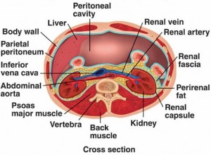
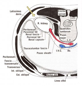
The kidneys have spaces in the surrounding areas that are divided into three distinct compartments, namely the perinephric, the anterior, and the posterior pararenal spaces. Besides these spaces, the renal fascia is surrounded by various attachments that connect various organs and vessels. The attachments are classified as below:
- Anterior attachment, which passes anterior to the kidney, renal vessels, inferior vena cava, and abdominal aorta. This attachment later fuses with the renal fascia’s anterior layer that is resent on the opposite side.
- Posterior attachment, which fuses with the vertebrae’s and psoas fascia
- Superior attachment, which consists of anterior and posterior layers, attaches at the kidney’s upper pole and then gets divided to enclose the adrenal gland. They fuse again at the upper portion of the adrenal gland to form the suspensory ligament of the gland and thereby fuse with the diaphragmatic fascia.
- Inferior attachment, which does not show the fusion of the layers but the posterior one moves downward and fuses with the iliac fascia while the anterior layer mixes with the connective tissue of the iliac fossa.
The anterior and posterior fascia blends to develop the lateroconal fascia that gets attached to the transverse fascia.
Renal Fascia – Clinical Relevance
The renal fascia is involved in various urinary tract diseases, including acute renal infection, intrarenal and perirenal masses, focal renal scarring, and renal calculi to name a few.
Renal fascia Images
Know how this urinary structure looks like with the aid of these pictures.
Picture 1 – Renal fascia
Picture 2 – Renal fascia Image
References:
http://en.wikipedia.org/wiki/Renal_fascia
http://www.wisegeek.com/what-is-the-renal-fascia.htm
http://anatomytopics.wordpress.com/tag/renal-fascia/
http://dictionary.reference.com/browse/renal+fascia


No comments yet.