Trochlear Nerve Definition
The Trochlear nerve is one of the smallest motor nerve (in terms of axons) that controls the superior oblique muscles (one of the muscles that helps in the movement of the eye) of the eye. The trochlear nerve is the fourth Cranial nerve having the longest intracranial course and is the only nerve that originates from the back of the brain stem. Damage to the trochlear nerves may result in the upward or side movement of the eyes often referred as vertical and torsional diplopia.
Characteristics of trochlear nerve:
- Smallest Nerve in terms of Number of Axons in our body.
- Travels Longest Intracranial route
- Only cranial nerve having a dorsal exit from the brain stem.
- Innervates Superior oblique muscles contralaterally, that is the opposite side from its origin.
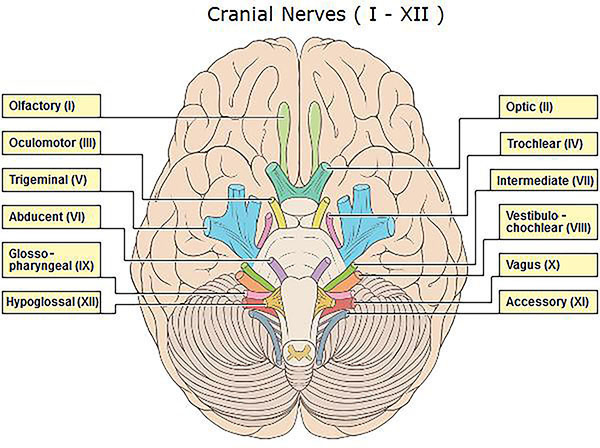
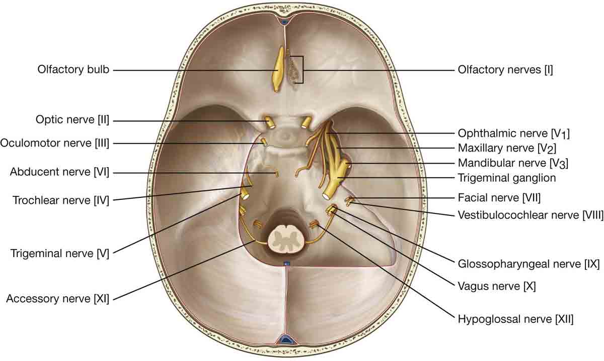
Trochlear Nerve Location and Origin
The trochlear nerves originate from the Trochlear nucleus. The trochlear nucleus is found on the posterior side of the midbrain. The exact location of the trochlear nucleus is deep below the fourth ventricle, towards the anterolateral side of the periaqueductal gray matter, just beside the inferior colliculus, located almost below the oculomotor nerve. The axons of the trochlear nucleus run dorsally around the aqueduct and further decussate immediately under the free margin of the tentorium cerebelli.
Trochlear Nerve Location
Course of Fourth Cranial Nerve
Each superior oblique muscle is provided with the nerve, from the trochlear nucleus, on the opposite side. This happens because as the nerve leaves from the trochlear nucleus, they immediately wind around the brain stem and cross to the other side of brainstem just before exiting from the brain stem. The fourth cranial nerve is supposed to be the longest intracranial nerve covering a length of approx. 60 mm.
The trochlear nerve after leaving the nucleus runs through the lateral side of the midbrain and enters into the cavernous sinus. The trochlear nerve enters the sinus through the anterior side always. Before entering the sinus, the trochlear nerve contralaterally travels through the cerebral peduncle, reaching and coursing through the midbrain. After moving around the midbrain, the trochlear nerve anteriorly runs below the posterior cerebral artery, while passing above the superior cerebellar artery, enters into the sinus. After reaching the sinus, the trochlear nerve moves along laterally through the sinus walls and reaches the orbit through the superior orbital fissure.
What is the function of the Trochlear Nerve?
The function of the trochlear nerve is to carry axons to the superior oblique muscles, which controls the movement of the eye. Trochlear nerves carry general somatic efferent types of the axon. The superior oblique muscles help in the movement of the eyes in the upward and downward movement. This is done with the help of Trochlear nerve axons, which are located on the medial aspect of the orbit. The superior oblique muscles pass through the loop of these trochlear nerves and help in oculatory movements.
Trochlear Nerve Pictures
Trochlear Nerve
Trochlear Nerve Picture
Clinical Implications
The trochlear nerve is often vulnerable to head injury or injury during surgery because of its small size and longest intracranial route. The injury to the trochlear nerve results in the loss of function of the superior oblique muscles, because of which the inferolateral ocular movement i.e. ability to look down and outward may be affected. The injury may cause the muscles to pull the back of the eyes, which in turn results in the front of the eye to move down and outward. This may cause diplopia or double vision. The two different types of diplopia are as below:
- Vertical Diplopia: In vertical diplopia, the injured muscles pull the eye little upward as compared to the normal eye position. Because of which the person, the person observes two visual field or two separate vertical images from each eye. Because of this reason, the patient has difficulty stepping downstairs, and often they tilt their head downwards to fuse the two images.
- Torsional diplopia: The trochlear nerves are also responsible for the sideways movement of the eyes. When we tilt our head to one side, the eyeballs automatically move laterally opposite side, so that vision does not change and vertical things appear vertical. However, injury in trochlear nerve may result in torsional diplopia, in which two different visual fields appear tilted with respect to each other. To overcome this the person may tilt his head to the opposite side to fuse the two images.
Causes of Lesions in Trochlear Nerve:
Injury to the trochlear nerve may be caused due to peripheral lesions or central lesions, both of which are described briefly:
- Peripheral Lesions: These are caused because of the damage in the nerve bundles. The damage in the nerve bundles may cause Acute palsy or chronic palsy. Acute palsy mostly results because of the head injury or trauma. The head injury may cause slight displacement of the brain stem. Patients with minor nerve damage may experience blurry vision, while major nerve damage may cause diplopia.
Chronic palsy, in general, is caused due to congenital damages in the nerve bundles. In these cases, the nerve bundles may not be fully formed or defective from birth. The symptoms may appear in early childhood or in some cases very late.
- Central Lesion: These are caused due to damage in the trochlear nucleus. The damage to the trochlear nerves or the axons within the brain stem may be caused due to hemorrhage, tumors, arteriovenous malformations, demyelination and few more.
No comments yet.