The upper extremity of the human beings has the largest number of bones. This part of the skeleton varies from being simple to complex. The various articulations and the different structures allow the multifarious movements of the hand. Amongst the parts of the upper extremity, the wrist is one of the complex parts in terms of structure and functionality. The wrist bone is biologically known as the carpal bones and is the connection between the fixed part and the hanging part of the arm.
The carpal bones differ in structure, function and in articulation also.
What are Carpal Bones and how many are there?
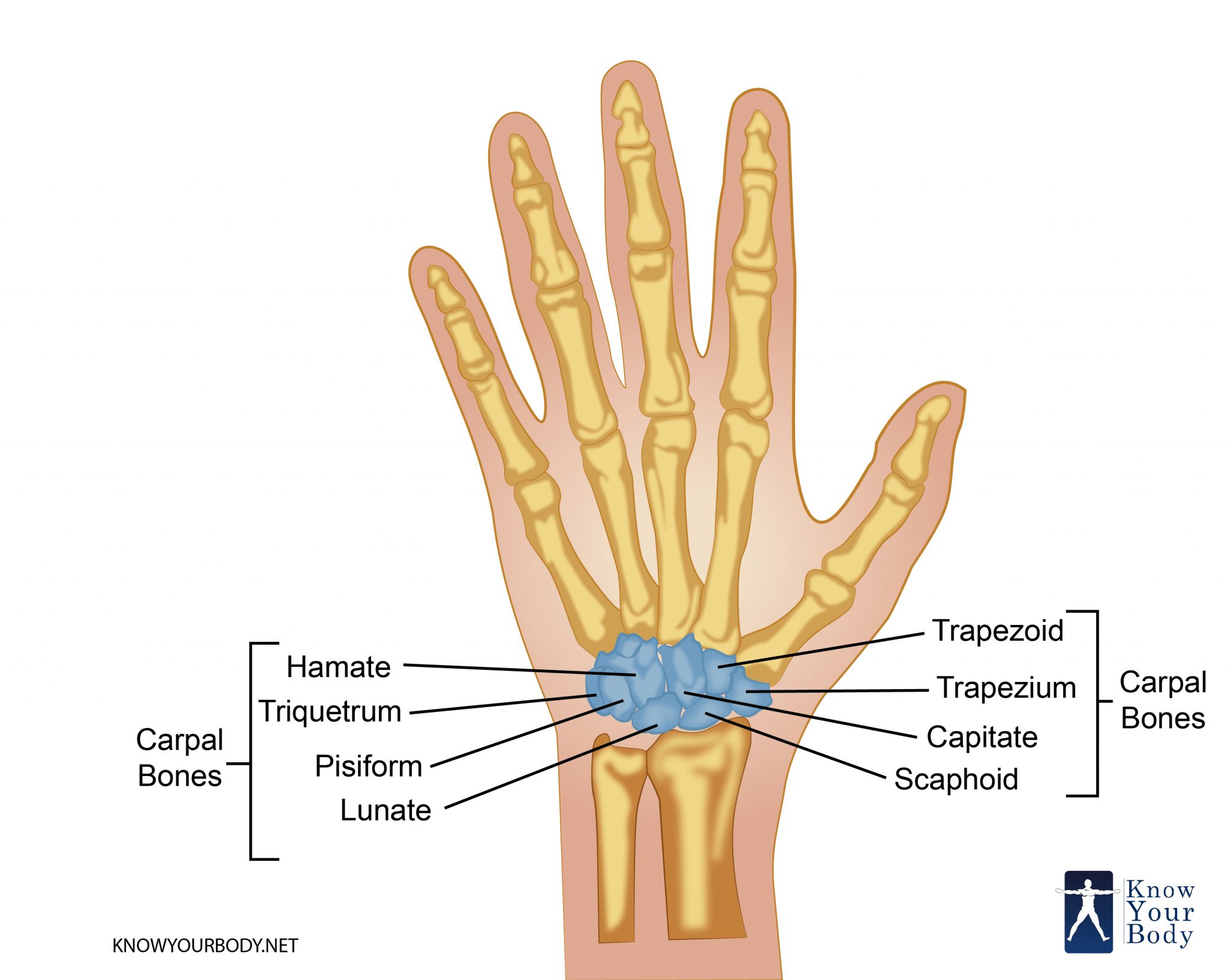
Wrist bone is the collective name of the carpal bones in our upper extremity. Eight bones form the juncture between the forearm and the hands. The eight bones in the wrist are named as:
- Scaphoid
- Lunate
- Triquetrum
- Pisiform
- Trapezium
- Trapezoid
- Capitate
- Hamate
These eight bones are arranged in the form of distal and proximal rows. Each row consists of four bones namely:
- The distal row consists of the trapezium, trapezoid, capitate, and hamate
- The proximal row consists of the scaphoid, lunate, triquetrum and pisiform
The transverse rows give the wrist its unique shape. The wrist bone is concave in shape in the distal region whereas the proximal surface shows convexity.
The eight bones can also be arranged in the form of longitudinal columns. They are three in number namely:
- The radial scaphoid column consists of the scaphoid, trapezium and the trapezoid
- The lunate column that consists of the lunate and capitates
- The ulnar triquetral column that comprises of the triquetrum and hamate
Location of the Carpal Bones
The carpal bone is located in the upper extremity of the human skeleton. The bones are the joining bridge between the ulna and radius and the metacarpals. The radius is articulated with the wrist bone on the proximal side. The metacarpals are articulated on the distal surface of the carpal bones.
Carpal Bones Location
The locations of the eight bones are as follows:
- The scaphoid is located at the radial border and is directed towards the thumb on the lateral side.
- Lunate is located at the articulation of the ulna and the radius. This bone occupies the central portion of the proximal carpal row.
- Triquetral is located on the ulnar side without any articulation with the ulna. This pyramidal bone is situated on the medial side of the proximal row.
- The pisiform is the last one of the proximal row and marks the ulnar border.
- The trapezium is situated above the scaphoid and forms the radial border of the distal transverse row.
- Trapezoid forms the second bone of the distal row and is almost punctured between the trapezium and capitate carpal bones.
- Capitates is the centrally located wrist bone and is often referred to as the wrist. It is situated on the distal surface of the ulna and the radius.
- Hamate marks the ulnar border of the distal transverse row.
Carpal Bones Structure
The structure of the eight carpal bones varies from one another, with having a different number of flat faces for the articulation of other bones.
Structure of Scaphoid
- Largest bone of the proximal row.
- A boat-shaped carpal bone that articulates with lunate, trapezoid, trapezium and the capitate carpal bone.
- In the proximal side, the scaphoid delineates convexity and articulates with the radius.
- The trapezium and trapezoid are articulated into the semi-lunar, distolateral facet of the scaphoid.
- The head of the capitate is articulated into the concave facet of the distomedial surface of the scaphoid.
- The lunate is articulated with the flat surface of the scaphoid bone.
- The scaphoid bone is narrow dorsally and having a groove for the attachments of ligaments.
Structure of lunate
- The lunate represents the crescent moon.
- It touches the radius on the proximal side.
- On the lateral side, it articulates into the flat surface of the scaphoid with a crescent-shaped facet.
- The medial face of the lunate bone is articulated with triquetral with a quadrilateral facet.
- Distally it is articulated with capitate.
- The lunate also shows articulation with the hamate on its distal and medial surface.
- The bone is concave on its distal surface whereas the dorsal side is rounded.
Structure of the triquetrum
- Three sided bone and hence is pyramidal in shape.
- The triquetrum is one of the carpal bone that forms the carpal arch.
- It articulates with the pisiform bone via an oval-shaped facet.
- The bone lies below the pisiform and hence is hidden mostly.
Structure of pisiform
- The pisiform is located at the ulnar border, at the juncture point of the ulna with the wrist.
- Only one articulation is found in pisiform where it articulates with the triquetral bone.
- Dorsally, the surface of the pisiform bone is oval in shape.
- The palmer side of the bone is rounded.
- The other two surfaces: lateral is concave in shape while the medial surface is convex.
Structure of trapezium
- On the ulnar side, it is surrounded with scaphoid and trapezoid bone.
- At the intercarpal joint, it is articulated with the trapezoid bone.
Structure of the trapezoid
- It contains four surfaces.
- The superior surface articulates with the scaphoid.
- The inferior surface articulates with the second metacarpal bone.
- The dorsal surface is larger than the ventral one and both provide area for attachment of the ligaments.
- Laterally, the surface is convex and is articulated with the trapezium.
- The capitate bone is articulated into the trapezoid medially.
Structure of the capitate
- Largest carpal bone amidst the eight bones.
- The hamate bone is articulated on the ulnar side while the trapezoid is located on its lateral surface.
- On the proximal end, it is surrounded by the scaphoid and lunate of the proximal transverse row.
- The distal or inferior surface of the bone has three facets being separated by two ridges. These three facets are articulated with the metacarpal bones of number II, III and IV.
- The ventral or palmer surface, support the attachment of the ligaments.
Structure of the hamate
- The hamate bone is connected to the metacarpal number IV and V.
- The narrow, superior surface of the wedged bone is articulated with lunate.
- The distal surface has two concave facets for the articulation of the metacarpals.
- In the palmar surface, the hamate has a hook-like structure known as the hamulus.
- The capitate is articulated to its lateral side.
Functions of the Carpal Bones
The functions of the carpal bones can be divided into two parts:
- LIGAMENT ATTACHMENT
The eight bones are connected with each other and with the other bones with the help of ligaments. These ligaments are divided into three groups:
- Ligaments of the wrist that connects the carpal bones with the radius and the ulna
- Ligaments of the inter-carpal articulations that joins the inter-carpal bones
- The carpals are joined with the metacarpals via the carpometacarpal ligaments
- AIDS IN MOVEMENT
- The vertical movement includes flexion and extension. Flexion is the downward movement of the wrist while the extension is the backward movement
- Rotation of the forearm consists of supination and pronation movements.
- Ulnar deviation is the movement of the carpal bones to the ulnar side while radial deviation shows the extension of the hand towards the lateral side
Carpal Bones Innervation
- The radiocarpal joint is innervated by the posterior interosseous nerves
- In the anterior portion of the carpal bones, the anterior interosseous nerve, median and ulnar nerves innervate the mid-carpal and the intercarpal joints
- The ulnar nerve and its deep-seated branch innervate the carpometacarpal joint.
Carpal Bones Clinical Significance
The common clinical signs that can be seen in all the eight bones are fractures and the development of osteoarthritis. However, there are certain specific diseases too associated with the carpal bones. They are:
- Scaphoid bone
- Scapholunate instability
- Preiser’s disease
- Lunate bone
- Kienbock’s disease
- Teisen classification
Carpal Bones FAQs
What are Carpal Bones?
The wrist comprises eight small bones referred as carpal bones along with two long bones located in the forearm known as radius and ulna. The carpal bone that is prone to injury the most include the scaphoid bone, lying in proximity to the thumb’s base.
What are the functions of the carpal bones?
The carpus constitutes the bony cluster of the wrist lying in between the metacarpus along with radius and ulna located in tetrapods. The carpus bones are never included in the fingers but they comprise the metacarpus bones. The part that constitutes foot part is known as Tarsus.
What are the muscles that remain attached to the carpal bones?
The muscle is known as the Flexor Retinaculum, which is an accessory ligament. It is made of the deep fascia lying at the wrist’s palmar surface. It remains attached hook of the hamate and the pisiform in a medial direction. It joins the trapezium and the scaphoid in a lateral manner.
What are the joints in the carpal bones?
The principal joints include plane joints, pivot joints, and hinge joints.
What is Carpal Tunnel Syndrome?
It is a disorder causing immense pain and numbness in the wrist and the hand. The symptoms arise while doing day-to-day activities like holding a telephone, driving a car and even reading a newspaper. The main reason for that dysfunction is due to the increase of pressure in the median nerve of the wrist.
No comments yet.