The hand is important for writing, grasping objects and doing our daily work. From outside, the hand might seem to be a single part of the body. However, anatomically, a hand is made up of different types of bones that bring about the motion by working in coordination.
There are twenty-five bones in the hand. Carpal bones, metacarpal bones, and phalanges are the three different types of bones present in the hand. Amongst these, the palm is made up of the metacarpal bones.
What are Metacarpal Bones?
Metacarpal bones are the intermediate skeletal structure of the hand that forms a connection between the carpal bones (wrist) and the phalanges (fingers). There are five metacarpal bones, numbered from I to V. Each metacarpal bone is connected to the proximal phalange of the concerned finger. The bone connected to the phalange of the thumb is numbered as I. The metacarpal bone corresponding to the little finger is the fifth bone.
Metacarpal Bones Location
The metacarpal bones are located in the intermediate position, between the phalanges and the carpal bones.
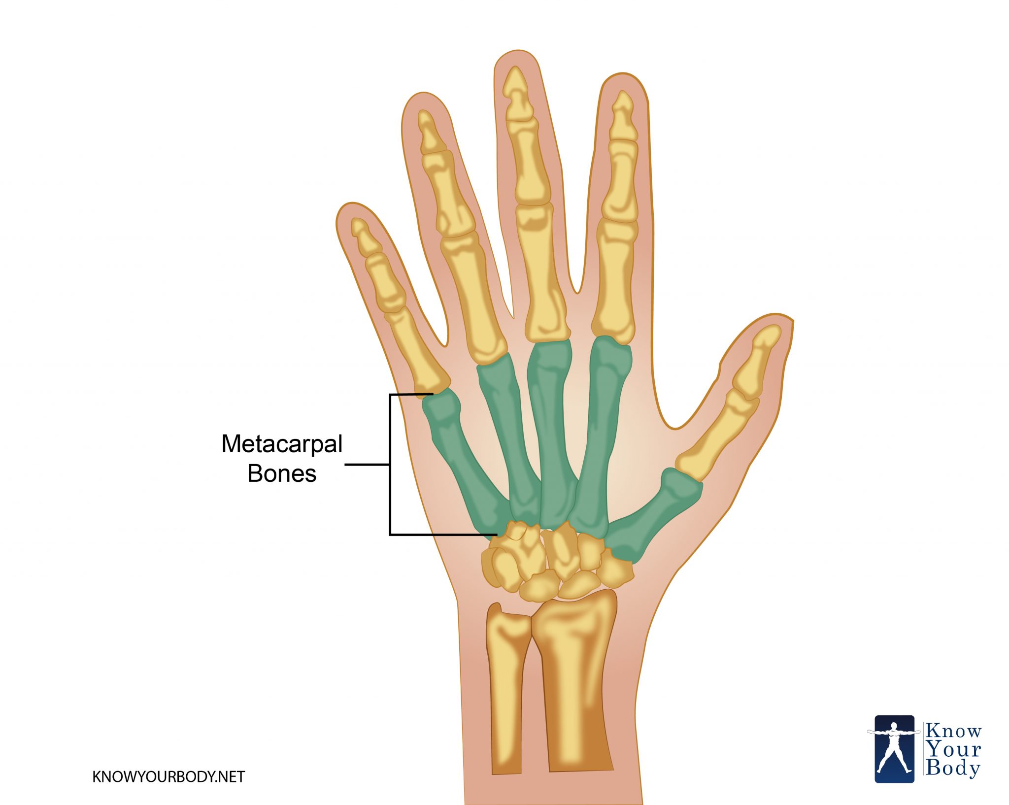
Metacarpal Bones Location
Metacarpal Bones Structure
The metacarpals bones are responsible for the concavity of the palm. When the two peripheral metacarpals i.e. the one attached to the thumb and the little finger, are brought close to each other, the concavity of the palm becomes prominent.
The independent movement of the five metacarpal bones is not same. The degree of fixation of the bones depends on the metacarpal concerned and can be described as:
- Metacarpal I or the thumb metacarpal is articulated with the scaphoid bone of the proximal transverse row of the carpals. This allows completely independent movement of the thumb.
- Metacarpal II or the index metacarpal is firmly fixed and it hence allows a considerable degree of movement.
- Metacarpal III or middle metacarpal is united with the carpal bone through intrinsic bone interlock. Hence, it’s movement is somewhat restricted.
- Ring metacarpal or metacarpal IV is a little bit mobile compared to the middle metacarpal.
- The last metacarpal bone i.e. the fifth one is partially independent.
Each metacarpal bone has a common structure:
- The distal extremity is known as the head and is in articulation with the proximal phalange of the digits.
- The head of the metacarpal bone or the digital extremity articulates with the proximal phalange of the concerned finger. The joint is termed as the metacarpophalangeal joint.
- The middle shaft is known as the body.
- The body or the shaft is prismoid in shape. The curve of the shaft is to introduce the concavity of the palm. Due to the resemblance with a prism, it has three surfaces: medial, dorsal and lateral.
- There is a transition region between the body and the head. This subcapital segment is known as the neck.
- The proximal extremity of the bone is termed as the base. It is connected to the carpal bones.
- The base is articulated with the carpal bones through the carpometacarpal joint and to the adjacent metacarpal bones also. The extremity is cuboidal in shape
The articulations of the metacarpals bones can be described as:
- Metacarpal I articulates with trapezium
- The second metacarpal is articulated with the trapezium, capitate, trapezoid and the third metacarpal
- The third or the middle metacarpal is articulated with the second and the fourth metacarpal.
- Metacarpal IV articulates with capitate, hamate, metacarpal III and metacarpal V.
- Metacarpal V articulates with the hamate and the fourth metacarpal.
Metacarpal Bones Function
The metacarpal bones act as the anchor for the phalanges and the carpal bones. The grasping ability of the Homo sapiens is due to the presence of these bones.
Metacarpal Bones Innervation
- The deep branch of the ulnar nerve supplies neural messages to the carpometacarpal joints. They also supply neural messages to the inter-carpal joints.
- The palmar digital nerves innervate the metacarpophalangeal joints. The dorsal digital nerves and the deep branch of the ulnar nerves also innervate these joints.
Metacarpal Bones Insertions
- Extensor Carpi Radialis Longus and Brevis: Base of the second metacarpal
- Extensor Carpi Ulnaris: Base of fifth metacarpal bone
- Abductor Pollicis Longus: Base of metacarpal I
- Opponens Pollicis: Thumb metacarpal
- Opponens digiti minimi: Inserts on the medial surface of the fifth metacarpal bone
Metacarpal Bones Clinical Significance
- Pseudohypoparathyroidism is a medical condition that is characterized by the shortening of the fifth and fourth metacarpal bones.
- A blunted Metacarpal IV and normal length fifth metacarpal define Turner syndrome.
- Nevoid basal cell carcinoma syndrome can also signify that the metacarpals are shortened in length.
- Boxer’s fracture usually occurs at the neck of the metacarpal bone.
Metacarpal Bones FAQs
What causes Boxer’s fracture?
This fracture is often seen in the neck of the metacarpal bones. Generally, the impact of a hard strike against a wall or any surface causes a crack in the metacarpal bones.
Do the boxers are prone to Boxer’s fracture only?
No, the boxers are not the only group of person who can get this injury. In fact, less trained brawlers are too at risk of fracturing their metacarpals.
What causes metacarpal pain?
A number of reasons cause pain in the metacarpals or precisely in the hands. Arthritis, fracture of the bones, etc are responsible for the aching of the hand.
No comments yet.