What is Occipitalis?
The Occipitalis muscle is a thin, quadrilateral muscle that arises by the tendinous fibers from lateral two-thirds of occipital bone’s superior nuchal line as well as from the temporal bone’s mastoid section.
Derived from Latin language, Occipitalis muscle is pronounced in Latin as Venter occipitalis musculi occipitofrontalis. Being one the skull covering muscle, occipitalis muscle is also known as the occipital belly of epicranius.
Occipitalis Location
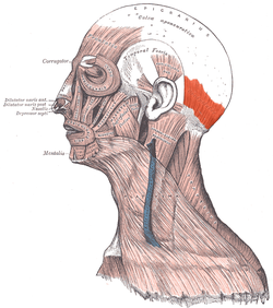
The muscle is positioned at the back of the head, above the base of the scalp, anteriorly surrounded by face and neck region.
Occipital Artery supplies blood to the occipitalis muscle.
Occipitalis Origin
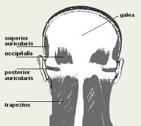
The muscle originates at the superior nuchal line,one among the four curved lines of the occipital bone, as well as the mastoid section of the temporal bone, bones present at the base and sides of the cranium. The muscle terminates in a tough dense fibrous tissue layer called the Aponeurotica’s Galea.
Occipitalis Description
The Occipitalis along with frontalis muscle is one of the two bellies or sections of the epicranius. The occipital belly lies close to the occipital bone. The muscle originates at the mastoid part of temporal bone as well as the occipital bone’s superior nuchal line and ends at the galea aponeurotica. It exchanges information with frontalis through an intermediate tendon. The muscle forms the posterior section of the occipitofrontalis muscle.
The flexibility of occipitalis muscle aids in the wiggling of the ears of many people.
The blood supply for occipital muscles is carried out by the occipital artery.
Occipitalis Function
Occipitalis Muscle is an important muscle responsible for facial movements. The muscle helps move the scalp and wrinkle the forehead as well as raise the eyebrows. The occipital section or the belly of the epicranius muscle helps an individual to extend the scalp such that the eyebrows may come up. It also helps to wrinkle the forehead.
Utilization of the complete muscle results in the closure of eyes by drawing the skin of forehead, cheek, and temple. Repeated such movements can cause a condition called Crow’s Feet, a condition characterized by the presence of branching wrinkle at the eye’s outer corner.
Occipitalis Problems
Tension or prolonged elevation of the eyebrows can cause pain to this muscle. The various health issues which may result from such pain include:
- Pain around the eyes or ocular strain
- Pain in the back and top region of the head which could be deep down in the head
- Tightening of scalp
- Migraines headaches
- Cluster headaches
- Tension headaches
- Temporomandibular Joint Dysfunction
- A toothache
It is noteworthy that the flexibility of this muscle helps wiggle the ears in many people.
Occipital Neuralgia
Occipital neuralgia is a condition characterized by shooting pain or paroxysmal stabbing in the dermatomes of the nervus occipitalis major along with nervus occipitalis minor. Pain may occur initially in the suboccipital area and then spread to the occipital and vertex region. Hypoesthesia and dysesthesia may also be present in the affected region.
Occipital Neuralgia Causes
Damage, trauma, or irritability of the nervus occipitalis major or minor due to causes occipital neuralgia due to injury or tightening of muscles around the nerves or inflammation.
Usually, secondary headaches are related to an underlying disease condition including hemorrhage, trauma, infection, tumor, and systemic disease.
Following conditions are associated with occipital neuralgia:
- Cervical disc disease
- Upper cervical spine’s osteoarthritis
- Tension or tightness of neck muscles
- Tumors impacting the C3 and C2 nerve roots
- Blood vessel inflammation
- Gout
- Infection
- Diabetes
Occipital Neuralgia Symptoms
Occipital neuralgia results in an intense pain accompanied with jabbing, sharp, and electric shock-like feeling in the posterior region of the neck and head. Following are the other symptoms which include:
- Neck ache
- Pain in either one or both regions of the head
- Sensitivity to light
- Eye pain
- Tenderness in the scalp
In certain people, the pain may be aching type rather than a jabbing, sharp or stabbing one. At the region where the occipital nerve passes in between the occipital and mastoid protuberance, there could be palpation and tenderness.
Occipital Neuralgia Diagnosis
Doctors will conduct an inquiry about the past medical history and injuries. Then a physical examination might also be carried out.
Sometimes the physician may administer medicine known as nerve block that numbs the nerves to analyze if it provides relief from pain. If it does, then the underlying cause of pain is most likely to be occipital neuralgia.
Apart from the above things, the following can be prescribed:
- Magnetic resonance imaging (MRI): Using computer technology and powerful magnets this test provides three-dimensional images of body components and hence can detect and reveal impingement of the spinal cord from disc, bone, or hematoma.
- Computed tomography scan (CT or CAT scan): In this diagnostic test, an image is provided after the computer reads x-rays and hence can reveal the size as well as the spinal canal, its components and the structures surrounding it.
- Blood test
Occipital Neuralgia Treatments
Non-surgical treatment modalities aim to alleviate the pain. Symptoms can disappear or improve with the following:
- Application of heat
- Rest
- Massaging strained neck muscles.
- Using anti-inflammatory medications such as ibuprofen or naproxen
- Use of muscle relaxants
- Taking antidepressants
- Taking oral anticonvulsant drugs such as gabapentin and carbamazepine
- Using percutaneous nerve blocks. These drugs reduce the pain apart from helping in the diagnosis of occipital neuralgia. Nerves which are affected includes occipital nerve, C2 and or C3ganglion nerves. Caution is advised while using nerve blocks as they contain which can cause adverse effects.
Surgery
Surgical intervention is not necessary unless there pain is chronic and extreme, and if there is no improvement even after using the above-said methods. Surgery may include:
- Occipital nerve stimulation: Electrical pulses are delivered to the occipital nerves using a device known as neurostimulator as they block pain reception in the brain.
- Microvascular decompression: Identification and managing the blood vessel causing nerve compression can help in reducing the pain.
Occipitalis Pictures
Here are some images that demonstrate the physical appearance of the muscle.
Picture 1 – Occipitalis
Picture 2 – Occipitalis Image
References:
http://en.wikipedia.org/wiki/Occipitalis
http://www.innerbody.com/image_musbov/musc66.html
http://wiki.answers.com/Q/What_does_the_Occipitalis_muscle_move


No comments yet.