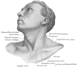
What is Suprasternal Notch?
It is defined as a large, prominent dip on the apex of the sternum in the middle of articulation along with two clavicles. It is a significant division of the human anatomy. In adults, the notch is mainly present due to an aortic arch aneurysm. In a child, it is present as a result of coarctation of the aorta.
Suprasternal Notch Synonyms
The structure is also referred to as:
- Fossa jugularis sternalis
- Jugular notch
Suprasternal Notch Location
It is located at the top edge of the manubrium right in the middle of the clavicular notches.
Suprasternal Notch Function
The Intrathoracic pressure is analyzed with the help of a transducer. In this process, the actuator employs the soft tissues which are situated right above the Jugular notch. A proper evaluative test for the aorta is conducted by utilizing this large dip. Such clinical tests can help determine a number of medical conditions such as:
- Dissecting aneurysm
- Hypertension
- Aneurysm
- Atherosclerosis
To proceed with such tests, it is compulsory to put an index finger or a middle finger right in the middle of the notch and tactually explore it. In average normal person, the palpable pulse is not present. If someone exerts more pressure in the Suprasternal Notch area, a palpable pulse can be felt, especially in older patients. Presence of a prominent pulse signifies the presence of uncoiled aorta.
Jugular Notch is used as a vulnerable spot for different finger strikes in martial arts such as Jiu-Jitsu. In this form of self-defense, an opponent may push their fingers right into this vulnerable spot in an attack known as “Two-finger strike.” This technique jams the windpipe and induces unconsciousness or even choking in a few cases.
People having problems in this notch are recommended to wear open neck garments at all times to make the region comfortable.
Suprasternal Notch Clinical Relevance
Palpation
The suprasternal notch gets palpated right in the mid-point of vital medial end points of the clavicles.
Epilepsy Treatment
If a patient suffers from epilepsy, drug diffusion takes place via the suprasternal notch or jugular notch. After anaesthesia, a 2 to 3 cm vertical incision is made in the inferior portion of the chin through the suprasternal notch. Once the sternothyroid muscle and the common carotid artery are exposed along with the posteriorly positioned vagal nerve separation, the drug is administered through catheterization, primarily in the form of a solution.
Suprasternal Notch Pictures
Take a look at these useful images to get an idea about the structural appearance of the notch.
Picture 1 – Suprasternal Notch
Picture 2 – Suprasternal Notch Image
References:
http://en.wikipedia.org/wiki/Suprasternal_notch
http://www.anesi.com/super.htm
http://www.meddean.luc.edu/lumen/meded/medicine/pulmonar/pd/pstep37.htm


No comments yet.