Petrotympanic fissure Description
It is a soft tissue or fissure located in the temporal bone which extends to the tympanic cavity from the temporomandibular joint. When running constantly with the petrotympanic fissure, both tissues are together often referred to as “Squamotympanic fissure”.
Petrotympanic fissure Names
This tissue is known by various other names like
- Glaserian fissure
- Fissura petrosquamosa
- Fissure pétrosquameuse
It is also known as “Fissura petrosquamosa” in Latin and “Fissure pétrosquameuse” in French. It owes the name “Glaserian fissure” to its discoverer, the famous anatomist Johann Glaser.
Petrotympanic fissure Location
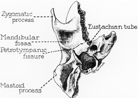
Picture 1 – Petrotympanic fissure
The tissue is located between the temporomandibular joint and the middle ear. The mandibular fossa, also known as Glenoid fossa, is bounded by the articular tubercle in front and by the tympanic part of the bone at the back. While the tympanic bone section splits it up from the external acoustic meatus the petrotympanic fissure divides the mandibular fossa into two parts and appears as a narrow slit.
Petrotympanic fissure Anatomy
The tissue opens just in front of and above the bony ring where the tympanic membrane is tucked in. In this position, it appears as a tiny slit that is about 2 mm. long. It houses the anterior malleus ligament and the anterior process. It also provides passage to the anterior tympanic branch positioned in the internal maxillary artery.
Petrotympanic Fissure Function
The fissure performs various functions:
- It allows communication between the middle ear and the temporomandibular joint (TMJ).
- It provides the tongue with special sensory (taste) innervation.
- It transmits Chorda tympani, anterior ligament of malleus as well as the anterior tympanic branch of the maxillary artery.
Petrotympanic fissure Contents
The fissure contains branches of cranial nerves VII as well as IX to the infratemporal fossa. A branch of the cranial nerve VII, known as the chorda tympani, goes through the fissure and joins the lingual nerve. The tympanic nerve communicates with the cranial nerve IX and passes as the lesser petrosal nerve through the fissure.
Petrotympanic Fissure Pictures
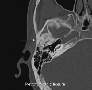
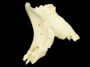
Here are some useful photos that analyze the structure of the tissue. Check these out to know how this temporal bone slit appears to view.
Picture 2 – Petrotympanic fissure Image
Picture 3 – Petrotympanic fissure Photo
References:
http://www.oluwoleogunranti.com/course/course1/miscellaneous/petrotympanicfissure.htm
http://www.rightdiagnosis.com/medical/petrotympanic_fissure.htm


No comments yet.