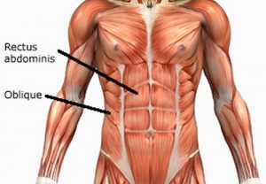
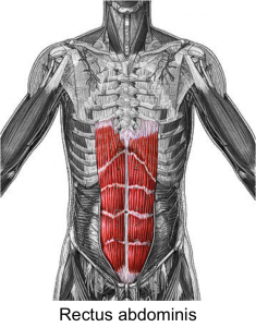
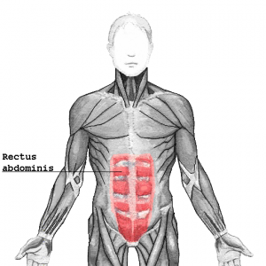
What is rectus abdominis and where is it located?
Rectus abdominis is the muscle that runs vertically adjacent to the umbilicus along the whole abdomen from the sternum up to the pubic bone. They are commonly known as abdominals or ‘abs’ muscles and are present in the pair at the front wall of the abdomen.
Rectus Abdominis Origin
The muscle is generally more developed in gym goers and athletes in whom the muscle is almost 20 mm thick rather than physically inactive individuals in whom the thickness is something around 10 mm. An opposite joint torque of the rectus abdominis muscle is created by its antagonist muscle erector spine.
Rectus Abdominis Insertion
From the inferomedial costal edge and back part of the xiphoid procedure of the sternum and fifth to seventh costal ligaments.
Rectus Abdominis Actions
Posteriorly angles with the pelvis region, Flexion of the vertebral segment, packs stomach substance, helps with strength termination.
Rectus Abdominis Innervation
Ventral Primary rami (T7 to T12)
Rectus Abdominis Structure
Rectus abdominis muscle is two flat and parallel muscles separated by linea alba (a connective tissue). Originating from the pubic crest (between pubic tubercle and pubic symphysis) at the lower end the muscle inserts with the help of fibers to the 5th to 7th coastal cartilage and the xiphoid process of the sternum. If the belly fat is burnt this muscle reveals the underlying 3-4 horizontal tendons intersecting the muscle. It results in the 4-6-8-10 packs although 6 packs are the most common. These intersecting tendons are the evidence of segmentation process during embryogenesis.
Rectus Abdominis Blood supply
Lower intercostal nerves along with its branches thoraco-abdominal nerves carries out the innervation process whereas the sensory stimulus relies on thoracic nerves. Blood is supplied to the rectus abdominis muscle through two arteries. Namely, an inferior epigastric artery supplying blood to the proximal part and the superior epigastric artery supplying blood to the distal part of the muscle.
Rectus Abdominis Functions
- Movement of the trunk.
- Flexing the spine.
- Stabilization of the vertebral column.
- Helps in respiration as an accessory muscle.
- Tensioning of the abdominal wall.
Rectus Abdominis Pictures
Rectus Abdominis Related Conditions
Abdominal hernias
The anterior abdominal walls or the rectus abdominis region are prone to hernias (i.e. abnormal protrusion of organ). Abdominal hernias show the following common characteristics like:
- Irreducible (it cannot be forced back to the abdominal cavity by pressure)
- Obstructed (blockage of the intestine leading to frank constipation and severe abdominal pain)
- Incarcerated (struck inside the abdominal cavity)
- Strangulated (loss of blood supply as the blood vessels get ruptured)
Hernias in this region are of varied types–
A spigelian hernia– Occurs when the linea semilunaris (the connective tissue separating rectum abdominis from lateral abdominis muscles) gets weak resulting the bowel to herniate through.
An umbilical hernia– It is common post-delivery or any other inter-abdominal pressure like ascites from the liver. It also occurs owing to the weakness of the posterior surface of the umbilicus, which in turn causes outpouching of bowels.
A paraumbilical hernia– When the bowel herniates through due to the weakness of muscles surrounding the umbilicus, this type of a hernia will form.
An epigastric hernia– This occurs when the musculature around the epigastric region (superior and inferior epigastric artery) gets weak.
An incisional hernia– This type of a hernia occurs post-surgical operations when the fascia (musculature) of the region is weak and the bowel herniates through.
Omphalocele/Exomphalos: It is a rare genetic disorder often associated with chromosomal aberrations like trisomy of chromosome 13 (Patau Syndrome) and trisomy of chromosome 18 (Edward’s syndrome). In this condition, as the abdominal layers fail to close, the abdominal content is held in a sac outside the abdominal cavity.
Gastroschisis
An incomplete development of the abdominal wall or if the rectus abdominis muscle fails to close during the fetal life the content of the abdominal cavity may herniate out. Unlike Omphalocele, Gastroschisis does not involve umbilical cord and actually occurs to the right side of the umbilical region. This disease could only be cured if the baby undergoes a surgical operation at a very early stage as the organs will swell and the cavity will shrink gradually as they grow, which will ultimately turn lethal.
Intercostal nerve Syndrome
Clinically also referred to as rectus abdominis syndrome, this disease refers to the trapping of the intercostal nerves to the abdomen supplemented with inflammation and rupture of the epigastric artery and stretching of rectus cutaneous medialis nerve.
Etiology
Although the definite cause of this disease is not discovered it is much noticed in women expecting a recent delivery, athletes, and labors.
Symptom
The symptom of rectus abdominal syndrome are the following-
- Chronic abdominal pain
- Acute abdominal pain
- Abdominal distension
- Mimic visceral diseases like appendicitis.
When the disease gets extreme then it starts to show signs like-
Bilateral pain in the upper thoracic region
- Precordial pain
- Nausea and vomiting
- Excessive and frequent abdominal cramp
- Colic
- Bloating
- Bilateral low back pain
- Swelling and gas
- intensified dysmenorrhea
Diagnosis
Being a rare disease and no such particular or defining external symptom except abdominal pain (which may occur due to several other reasons) and also mimicking symptoms of other diseases the disease remains undiagnosed in 80-90% cases. Lidocaine test is the only test to diagnose the disease. The process includes injecting lidocaine into the subcutaneous layer of the sheath of rectum abdominis near the rectus cutaneous medical nerve.
Treatment
Following are the most common treatments that are done to treat this disease
- Local steroid injection
- Local anesthetic surgery
- Spinal cord stimulation
Hematomas
Here is another uncommon disease on the list. While hematomas (clotting of blood to solid structures within tissues) are more common in organs like liver, kidney, heart etc., rectus abdominis hematoma, popular known as Rectus Sheath Hematoma (RSH) in medicine practitioners is one of the most severe disease in the lost since like other hematoma it is an emergency condition. While post-surgical or post-delivery hematoma is readily checked but when the same causes due to bleeding from a ruptured epigastric artery or direct tearing of rectus muscle, the condition goes undiagnosed as the symptoms mimic like any other abdominal pain. In such misdiagnosed and hence mistreated condition the affected individual will dive to the lap of death within a few hours.
Rectus abdominis is one of the largest single muscle in our body and its evolutionary significance in maintaining the proper posture of our body cannot be unaccepted. It also adds to the physical aesthetics when the individual works on their abs. But we should also be committed to our health and must take special care that we do not use these muscles excessively leading to a severe disorder.
References:
https://www.kenhub.com/en/library/anatomy/rectus-abdominis-muscle
https://www.ncbi.nlm.nih.gov/pubmed/3160269
http://reference.medscape.com/article/776871-overview
http://www.rightdiagnosis.com/medical/rectus_abdominis_syndrome.htm
http://www.webmanmed.com/disorders/disorders_files/musclgd/abdom/12283745.html
http://www.healthline.com/human-body-maps/rectus-abdominis-muscle


No comments yet.