What is the Sphenoid Bone?
The sphenoid bone, also known as Os sphenoidale, is a cranial bone, shaped like a butterfly or a wasp, which occupies most of the middle part of the skull’s base, making up the middle region of the cranial fossa. Crammed between a group of bones in the frontal part of the cranium, it becomes one of the 8 bones that form the cranium. The sphenoid bone primarily consists of a central body (containing the pituitary gland) and two pairs of wing-shaped extensions projecting laterally. Forming the sides and floor for eye orbits, it keeps the eyes intact.
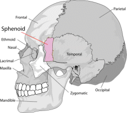
Where is the Sphenoid Bone located?
The sphenoid bone makes the floor of the skull, present in front of the temporal bones and making the base of the occipital bone. It is segregated into a middle part called the body, a pair of great wings and a pair of lesser wings.

Sphenoid Bone Location
What is the Gross Anatomical Structure of the Sphenoid Bone?
Being the most complex structured bone of the human skeleton, it is divided into the following regions:
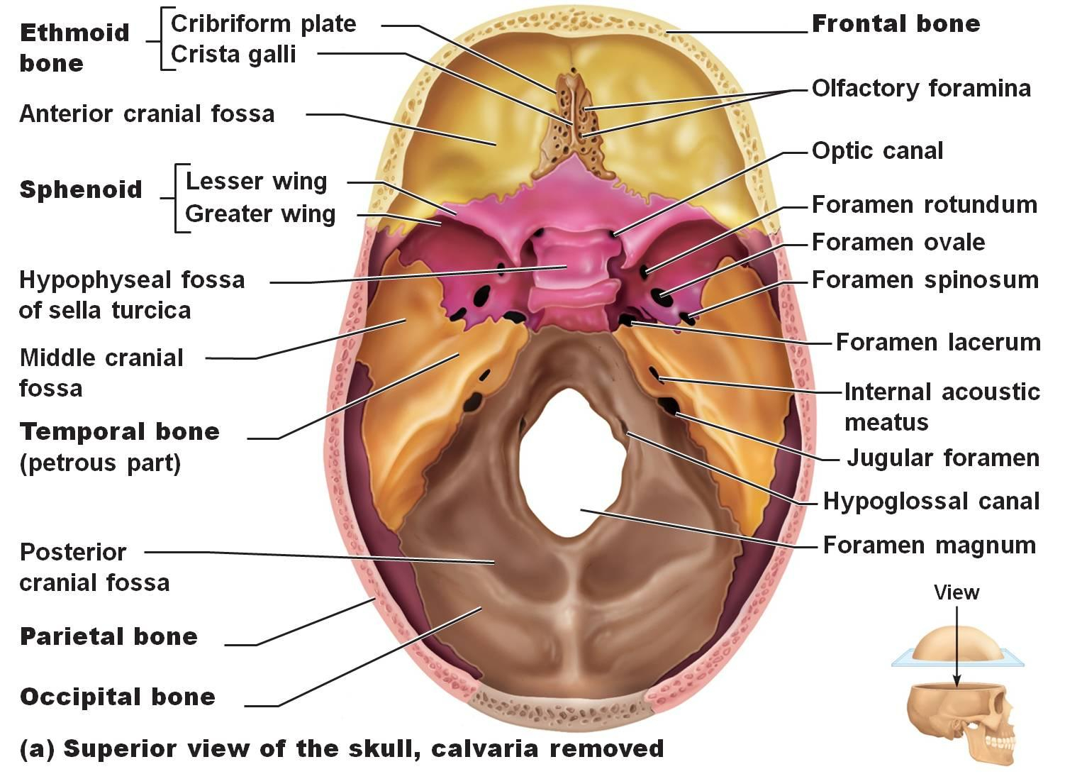
- Body is a nearly cubical-shaped bone creates the middle part of the sphenoid bone called the body or the corpus shenoidale. It consists of two sphenoidal air sinuses, separated with the help of a bony septum. This part shows articulation with the ethmoid bone in the anterior region, which lets the sinuses open into the nasal cavity.
- The body’s superior surface is located ventrally to an important spine, called the ethmoidal spine and articulates with a particular cribriform plate (part of the ethmoid bone).
- On the back side of this is a comparatively smooth surface with a highlighted midline, prominent on both sides of the cerebrum’s olfactory lobes.
- Sella turcica is a depression which can be seen as a saddle and constitutes 3 parts:
- Tuberculum sellae – the anterior surface of sella turcica and posterior part of groove named chasmatic groove.
- Hypophyseal fossa – deepest hollow space of sella turcica where the pituitary gland is situated.
- Dorsum sellae – the wall of sella turcica lying posterior.
- Chiasmatic Groove is a groove formed due to the crossover of the optic nerves known as the optical chiasma.
- A part of anterior, as well as posterior Clinoid Processes, encompass the sella turcica. Anterior ones arising from the lesser wings of the sphenoid bone and the posterior ones form the superiolateral processes of the dorsum sellae. They are the points of attachment of a membrane dividing the brain called the tentorium cerebelli.
- The canalis opticus or the Optical Canal opens near the lesser wings of the sphenoid bone and functions as a tunnel for the optic nerve as well as the ophthalmic artery to pass.
- Lesser wings, also known as Ala minor are a pair of the smaller wing-like projections of the sphenoid bone. They are flat, triangular structures situated anterior to the greater wings.
- Greater wings, also known as Ala major is a pair of larger winged structures of the bone, curved upwards, backward and on the lateral sides to form a definite cavity or fossa in between the floor of the cranium.
- Foramen Ovale is an ovular hole present in the posterior region of the area of the greater wing. It forms a passage for a branch of the trigeminal nerve called the mandibular nerve as well as the lesser petrosal nerve, emissary veins and the accessory meningeal artery.
- Foramen Spinosum, a small orifice passing through it, the middle meningeal artery and the meningeal branches that are formed by the mandibular nerve, lies in the posterior region of the foramen ovale.
- Foramen Rotundum is another orifice situated both anteriorly and in the middle of foramen ovale and helps in passing the maxillary nerve branching from the trigeminal nerve.
- Pterygoid Process is a protrusion descending from the junction of the greater wing and the sphenoid body. It is made up of two vertically oriented, thin plates, fused at the anterior end and separated by a cleft at the posterior end.
- The Lateral Pterygoid Process forms a point of anchorage for the lateral pterygoid muscles, which help in the mandibular movement during chewing action.
- The Median Pterygoid Process is the middle plate which is a longer and narrower one. It attaches the superior lying pharyngeal constrictor muscles and also the tensor veli palatine muscles, which aid in the process of swallowing.
- Pterygoid Hamulus is a hook-shaped low situated process of the medial pterygoid process. It enables gliding action of tendon associated with tensor veli palatine muscle and aids in tensing the palate during the act of swallowing.
- Articulations: the sphenoid bone can be seen articulated with other twelve bones which are, four singular bones namely, ethmoid, frontal, vomer and occipital, and four pairs of bones which are parietal bones, zygomatic bones, temporal bones and palatine bones.
Sphenoid Bone Functions
The sphenoid bone mainly plays a key role in the development of important anatomical structures and also, in the formation of the skull. Other functions are:
- There are many oval or circular foramina/orifices through which arteries and nerves of the head and neck region pass through.
- The sphenoid bone is a perfect junction for attachment of muscles which help in chewing food.
- The development of the skull cap and the lateral sections of the fossa are assisted by the sphenoid bone.
Sphenoid Bone Pictures
What is Transsphenoidal Surgery?
The sphenoidal bone gains its significance as it encases the pituitary gland, also known as the master endocrine gland, within it. Hence, it becomes easily possible to access the pituitary gland during surgery by the passage of the sphenoid bone and its sinuses. When an instrument is passed as described above, for treatment of various pituitary anomalies and adenomas, the surgical process is known as endoscopic trans-sphenoidal surgery or ETSS. This does the needful without going into a detailed craniotomy.
No comments yet.