Cecum Definition
Cecum or also known as “caecum,” is a small intraperitoneal pouch-like structure located on the right side of the lower abdomen. It is the starting point of the large intestine and lies between ileum and the ascending colon, into which the ileum (last part of the small intestine) empties digested content from the small intestine, to be further processed and digested in the large intestine.
Cecum helps in absorbing water from the solid waste produced by the small intestine and also lubricates and further processes the waste with the help of various enzymes to be digested in the large intestine and through the end stage of digestion.
Cecum Location
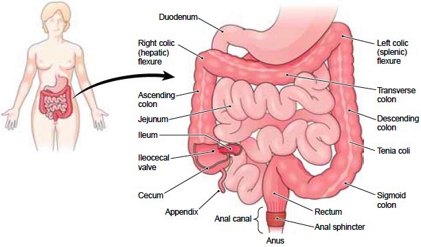
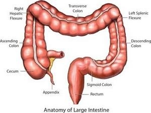
The cecum is a small sac-like structure at the beginning of the large intestine. To be precise, the cecum is the most proximal part of the large bowel and is located in the lower quadrant of the abdominal cavity on the right-hand side and usually lies laterally beside the ileum. In normal conditions, the cecum is approximately 6 cms in length with a diameter of 9 cms.
Cecum is located just below the ileocecal valve and may be felt in the right iliac fossa and the suprapubic region.
Cecum Location
Histology and anatomy of the Cecum
The cecum comprises of four different layers of tissue all having distinct functions:
Mucosal Layer: The mucosal layer forms the innermost layer of the cecum, comprising of mucous membrane containing various goblet cells. The goblet cells are responsible for releasing mucus, which helps in lubricating the cecum walls. The surface of the mucosal membrane consists of epithelial cells which help in the absorption of the nutrients from the undigested food.
Submucosal Layer: The mucosal layer is surrounded by the submucosal layer which consists of the blood vessels and nerve cells that help the intestinal tissues.
Muscularis: The third layer comprises of the crisscross bands of smooth muscle fibers known as the muscularis. The smooth muscle fibers help in contraction of the cecum walls which in turn helps in the churning of the semi-digested food and mixing with mucus and essential enzymes.
Serosa: Simple squamous epithelial tissue forms the outermost layer of the cecum, also known as serosa. This layer helps in lubricating the cecum and prevents it from frictional injury with the nearby tissues.
Function of the Cecum
The cecum is supposed to be the first part of the large intestine where the digested food enters from the small intestine. Partially digested food known as chyme enters into the cecum through the ileocecal sphincter, which opens and closes at regular intervals. Chyme inside the cecum is mixed with various bacterial enzymes and mucus before pushing it towards the ascending colon.
Various functions performed by cecum are Liquid Receptacle, Absorbing Salts, lubrication and Cellulose Breakdown.
Liquid Receptacle: Cecum acts as a liquid receptacle. The small intestine absorbs all the nutrients from the food and transfers the solid and liquid waste towards the large intestine. The cecum is the first part of the large intestine helps in receiving the liquid waste and helps in absorbing liquid from the wastage.
Absorbing Salts: Apart from being a reservoir for liquid waste the cecum also helps in absorbing the salts and electrolytes from the liquid food waste. There is a continuous loss of salts and electrolytes from the body while sweating. The contraction in the cecum muscles helps in the churning of the liquid waste food, while the mucous membrane of the cecum wall, in turn, helps in absorbing the salts and electrolytes and helps in replenishing our body of lost salts and minerals.
Lubrication: The lining of the cecum has numerous mucous producing cells. The cells help in lubricating and transferring of the solid waste through the large intestine. The large intestine absorbs water from the solid waste which makes the solid waste to move further. Lubrication of the solid waste by mucus walls of cecum helps in the movement of the solid waste through rest of the large intestine.
Cellulose Breakdown: Cecum also has essential cellulose-digesting enzymes, which help in breaking the cellulose fibers. The enzymes in the cecum help in fermentation and breaking down of cellulose fibers so that it can be easily digested later in the large intestine. Thus, cecum forms an essential part of the digestive system for many plant-eating herbivore animals and is found to be bigger in size as compared to omnivores, where cecum is very small and non-essential.
Cecum Pictures
Cecum

Cecum Picture
Disorders associated with Cecum
The large intestine comprises of five different segments, cecum being the first part of the large intestine. The important function of the large intestine is to absorb water, salt, and minerals from the digested solid waste. The disorders may develop in any part of the large intestine including cecum. Few of the important disorders associated with the cecum and various other parts of large intestines are as below:
Crohn’s Disease or IBD: This is an inflammatory bowel disease which is suspected to be caused due to a weak immune system triggered by a severe bacterial infection. Crohn’s disease is found to be associated with severe diarrhea, watery stool, pain in the abdomen with decreased appetite along with fever. The infection may be localized or occurs in patches and can affect cecum. In general, inflammatory bowel disease symptoms may not be regular and show up again after irregular time intervals.
Ulcerative Colitis: The symptoms of ulcerative colitis are similar to that of Crohn’s disease and include abdominal pain, watery stool, and diarrhea, vomiting, low appetite, tiredness, and anemia. This is also a kind of inflammatory bowel disease and an inflammation of the inner lining of the large intestine is the main characteristic of this disorder. The infection normally starts from the rectum and starts affecting the tissue lining upwards through the colon and reaching upwards to the cecum. In Ulcerative colitis patients, the tissue lining the large intestine starts dying causing the formation of ulcers which produce pus and causing blood in the stool.
Diverticulitis: Diverticulitis is characterized by the formation of pouches or small sac-like structure on the intestinal linings. These small pouches are called as diverticula, the presence of a large number of pouches results in a condition known as diverticulosis. Diverticulitis is caused when the food trapped inside the pouches starts getting decaying and causes inflammation of the intestinal linings. The characteristic symptoms associated with diverticulitis are fever with chills, vomiting, diarrhea, and sharp pain in the abdomen. In severe conditions this may cause intestinal bleeding, blocking of the colon, tearing of intestinal tissues etc.
Colorectal Cancer: The exact cause of colorectal cancer is unknown, however disorders associated with a colon like Crohn’s disease, polyps, history of ulcers may increase the possibility of colorectal cancer. The general symptoms associated with colorectal cancers are blood in stool, loss of appetite, incomplete stool elimination, the regular urge of defecating, bloating, sudden weight loss and discomfort or pain in the abdomen. Colorectal cancer may affect any part of the small or large intestine and specifically can affect cecum as well. Depending on the seriousness of the disease the doctor may suggest surgery and adjuvant therapies like chemotherapy and radiation.
Cecal Volvulus: This is a condition where there is an axial rotation of the cecum also involving the terminal ileum and some part of the ascending colon. In some cases, the rotation may be an upward and anterior direction of the ascending colon instead of the axial rotation and is referred to as Cecal Bascule. Both the conditions are distinct from each other however, both conditions result in the blockage and strangulation of the large intestine. This, in general, is caused due to the congenital defects in the intestinal formation during the fetal development. The symptoms associated with these disorders are a dark starry stool, abdominal pain, vomiting, abdominal distention and severe constipation.
FAQs
What is the difference between cecum and appendix?
The cecum is a sac or pouch-like structure located in the lower fourth quadrant of the abdomen on the right side. The appendix also known as a cecal appendix or vermix is a 2-20 cm blind-ended tube attached to the cecum near the hip bone. An appendix is normally considered as a vestigial organ.
What causes a twisted cecum?
The twisted cecum is a very rare condition and is mostly congenital, i.e. caused due to the weakness of the abdomen muscles since birth. The cecum is the starting point of the large intestine and is attached to the abdomen through a connection called mesentery. In certain people, the connection may be long mesentery or an incomplete attachment, which makes the cecum lose or “floppy” and resulting in twisting of the cecum. This condition is often referred to as “volvulus”.
What is inflammation in cecum?
Inflammation in cecum is caused due to severe diarrhea caused due to bacterial infections like Clostridium difficile. Neutropenic enterocolitis also is known as typhlitis is known as inflammation of cecum and is caused due to intestinal diseases and severe bacterial infection and is associated with lower levels of neutrophil count in the blood. Typhilitis is mostly found to be associated with patients who have undergone chemotherapy, AIDs, kidney transplant or elderly people who have a weak immune system.
No comments yet.