The digestive system is involved in the breakdown of complex food into smaller absorbable form. The ultimate fate of the food we eat is cellular respiration. The form in which the food is present is not absorbable by the cells. That is why digestive system breaks them into their ultimate product, which is diffusible through the cell membrane.
Digestion occurs in many sites of the entire alimentary canal. The last site of digestion is the small intestine. Furthermore, the small intestine is also the site of absorption of the digested nutrients into the bloodstream.
Small Intestine Definition
The small intestine is a highly coiled tubular structure that forms the end site of digestion. It is the most important part of the alimentary canal and leads to the large intestine. Sometimes, this organ is also called ‘small bowel’.
The ‘small’ term is added because it is shorter in diameter as compared to the large intestine.
Where is the Small Intestine located?
The small intestine sits in the lower abdominal region. The tubular structure starts from the pylorus sphincter of the stomach. It then coils around itself and leads to the opening of the large intestine, caecum. It covers most part of the middle and the lower section of the abdomen.
Small Intestine Location and Anatomy
At the junction point, there is a small pouch-like structure called the vermiform appendix. This is a vestigial organ in human beings and hence it is functionless.
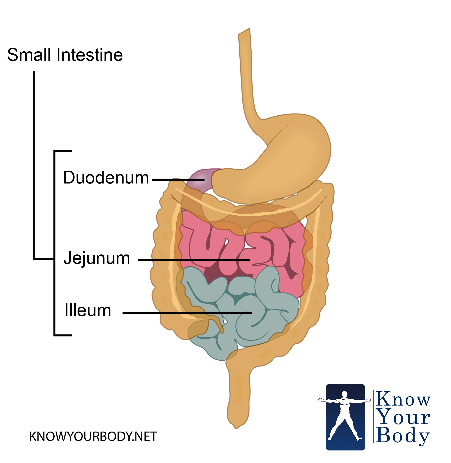
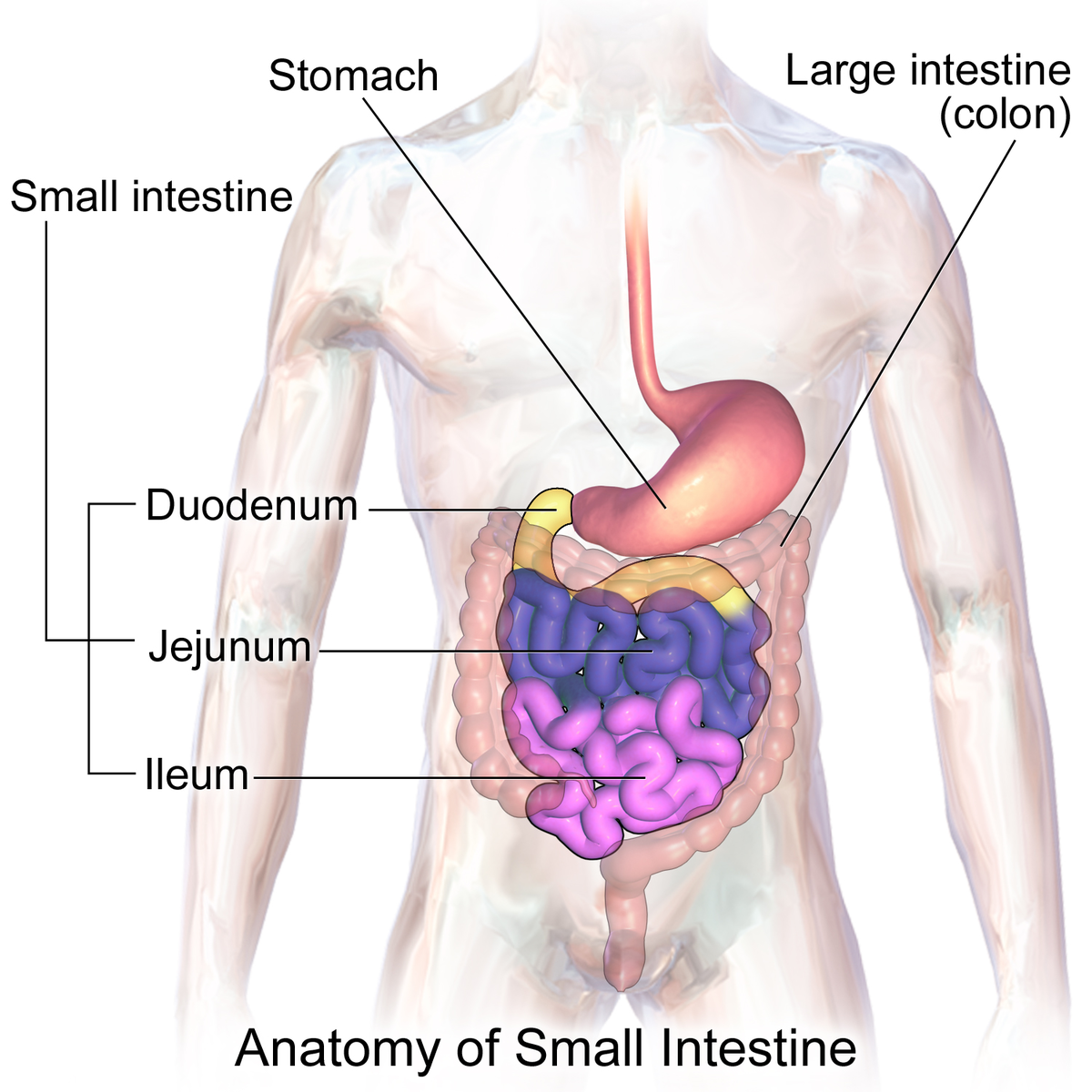
Parts of the Small Intestine
The small intestine is divided into three major parts, each characterized by its function and length.
The parts of the small intestine are:
The starting part of the intestine is known as the duodenum.
Duodenum leads to the middle and small length jejunum.
The jejunum is followed by the longest and the most coiled part of the intestine, the ileum.
Ileum leads to the large intestine through caecum.
How long is the small intestine?
The small intestine is exceptionally large, ranging 3-5 meter in length in humans. However, the length varies from person to person. More the height of the person more will be the length of the intestine.
The approximate diameter of the coiled structure is around 1.5 centimeter. However, this length can increase in case of dilation of the bowel.
Structure of the Small Intestine
The surface area of the small intestine is largely increased because of:
- The highly coiled structure allows a greater surface area
- The presence of microvilli in the ileum
Structure of Duodenum
- Duodenum is the first part of the small intestine that is connected to the stomach through the pylorus.
- The C-shaped section surrounds the head of the pancreas and receives the acidic chyme from the stomach.
- The entire length of the duodenum is divided four subparts- superior part, descending part, horizontal part and ascending part
- The superior part starts from the pylorus and ends in the superior duodenal flexure. It sits at the level of the first lumbar vertebrae and is mobile in nature.
- The descending duodenum lies between the superior and the inferior duodenal flexure. This part of the duodenum receives the common bile duct and the pancreatic duct through perforations in the region of the major duodenal papilla.
- The horizontal part passes over the inferior vena cava and the abdominal aorta. Sometimes, this part is also referred to as inferior duodenum.
- The final part, ascending part of the duodenum terminates at the duodenojejunal junction from where it leads to the jejunum.
Structure of the jejunum
- The jejunum is longer than the duodenum in length while shorter than the ileum.
- The inner mucosa of the jejunum is thrown into folds called villi.
- The walls of the jejunum have radial and longitudinal muscle which brings about the peristaltic movement.
- It is suspended from the abdominal section through the mesentery.
Structure of the ileum
- The longest and the most coiled part of the small intestine is the ileum.
- The inner mucosa membrane of the ileum is also folded into finger-like projections called villi.
- The radial and the longitudinal muscles help in the movement of the food along the intestinal tract through peristalsis.
- The ileum terminates at the ileocecal valve, the separation between ileum and caecum.
- There are numerous lymphatic patches distributed in the ileum and known as Peyer’s patches. These are areas where no villi are present.
Small Intestine Histology
There are four layers of the small intestinal wall, each different in the three parts of the intestine. The four layers are:
- Mucosa
- Submucosa
- Muscularis Externa
- Serosa
Mucosa layer
- The mucosa is the innermost layer surrounding the lumen of the small intestine. It is secretory, protective and absorptive in nature. The mucosa is further divided into three sublayers:
- Intestinal epithelium: the immediate epithelial layer surrounding the lumen consists of the micro projections villi and circular folds. Both these structures increase the surface area of the intestinal lumen. This layer contains five types of cells which have their own functions.
- Lamina propria: the second layer of the mucosa which is concerned with secreting mucus into the lumen of the tube through ducts. It consists of loose connective tissue that acts as a binding factor for the intestinal epithelium and the best part of the wall.
- Muscularis mucosae: third and the end sub-layer of the mucosa and is made up of smooth muscle fibers. This layer separates the submucosa from the lamina propria.
- The five types of cells in the mucosa layer are as follows:
- Nutrient absorbing cells
- Protective cells that secrete mucus into the lumen
- Hormone-secreting cells that secrete hormones like secretin, pancreozymin, etc.
- Anti-microbial cells or Paneth cells that are concerned with the secretion of human beta-defensin, a type of anti-microbial peptides.
- Immune cells that secrete lymphocytes into the lumen.
Submucosa layer
- The second layer of the intestinal wall is the submucosa layer.
- It is a thin layer that is made up of connective tissue
- This layer serves as a pathway for the vascular bundles and the nerves to enter the mucosa layer of the intestine.
Muscularis Externa
- Composed of two muscular fibrous layers and lies next to the submucosa layer
- The inner layer consists of circular muscle rings while the external layer consists of the longitudinal muscle fibers.
- This layer is responsible for generating the peristaltic contraction and relaxation and help in the movement of the luminal content.
Serosa layer
- The mesothelium layer is divided into two protectives covering: the inner viscera and the outer parietal layer.
- The gap between the layers is filled with connective tissue
- The secretion of the layer is known as serous fluid. This acts as a lubricant that allows frictionless movement of the muscles.
Small Intestine Functions
The small intestine is not concerned with only digestion. Rather it shows various functions that include:
Role in Digestion
Duodenum and ileum are the two sites of digestion.
- Duodenum receives bile from the common bile duct. This juice helps in turning the medium into alkaline. Also, it helps in the emulsification of the fats into small droplets of fatty acids.
Bile
Fats – emulsified fats
- The pancreatic juices are poured into the duodenum via the pancreatic duct. These juices act on the acidic chyme and break the complex nutrients.
Pancreatic amylase
Carbohydrate – maltose
enterokinase
Trypsinogen – trypsin
trypsin
Proteins – peptides+peptone
Pancreatic lipase
Emulsifies fats – fatty acids+glycerol
- These contents pass through the jejunum where no digestion takes place. The final site of digestion is the ileum. Here the intestinal walls secrete the enzymes.
maltase
Maltose – glucose
lactase
Lactose – glucose+galactose
sucrase
Sucrose – glucose+fructose
erepsin
Peptides+peptones – amino acids
lipase
Emulsified fats – fatty acids+ glycerol
Role in Absorption
The absorption of the digested products is done through the finger-like micro-projections, villi. The inner mucosa membrane is convoluted in folds that increase the surface area for the absorption. Half of the absorption occurs in jejunum and the leftover products are done in the ileum.
- The blood capillaries within the villi absorb the glucose and amino acids. Then they are taken to the liver via the hepatic portal vein for assimilation.
- The fatty acids are glycerol are absorbed through the lacteal duct which joins together and forms the lymph vessel. These end products are poured into the main bloodstream near the carotid artery.
- The villi are single layer thick structure. This facilitates the diffusion of the molecules into the bloodstream against the concentration gradient.
- The duodenal walls absorb iron while Vitamin B12 and bile salts are absorbed in the ileum.
- Water is absorbed throughout the length of the tubular structure
- Sodium bicarbonate salts are absorbed by active transport and hence it requires energy
Immunological Role
- The Peyer’s patch contributes to the production of lymphocytes, which helps in maintaining the immunological ambiance of the intestine.
Innervations of the Small Intestine
The autonomous nervous system i.e. sympathetic and parasympathetic nerves trigger the neural functioning of the small intestine.
- The vagus nerve branches off into parasympathetic fibers and enters the duodenum through superior and the celiac mesenteric plexus.
- The sympathetic nerves enter through the intestinal plexus.
- The same nervous system innervates the rest of the small intestine through the mesenteric plexus.
Small Intestine Pictures
Small Intestine Diagram
Clinical Significance of Small Intestine
A number of clinical conditions harm the small intestine. Starting from physical obstruction of the lumen to bacterial or viral symptoms, there are many complex disorders.
Physical obstructive disorders
- Hernia
- Adhesions
- Paralytic ileus
- Volvulus
Infectious diseases
- Ascariasis
- Tropical sprue
- Hookworm
- Nematodes
- Protozoan infection
- Giardiasis
Bacterial infections
- Cholera
- Dysentery
- Diarrhea
- Infections caused by Mycobacterium, Campylobacter, Shigella, etc
- Typhoid and paratyphoid fever
- Botulism
Viral infections
- Adenoviral infection
- Rotaviral infection
- Astroviral infection
Neoplastic disorders
- Adenocarcinoma
- Carcinoid
- Gastro-intestinal stromal tumor
- Sarcoma
- Melanoma
Genetic disorders
- Meckel’s diverticulum
- Pyloric stenosis
- Pancreas divisum
- Situs Inversus
- Cystic fibrosis
- Primary bile acid malabsorption
- Gardner syndrome
FAQs
What is malabsorption?
This is a clinical condition which is characterized by the improper absorption of nutrients by the small intestine. It can occur due to a number of reasons and one should consult a doctor for the further diagnosis.
What is gastroesophageal reflux?
Gastroesophageal reflux is a condition where the oesophageal sphincter muscle does not close properly and the food is pushed back into the esophagus along with the gastric juice.
What do you mean by celiac disease?
Celiac or gluten allergy is a condition where the protein called gluten triggers the immune response of the body. The immunological cells attack the small intestine, thereby causing pain, discomfort, and malabsorption.
What is endoscopy?
Endoscopy is a method of diagnosis where a small diameter pipe is inserted through the pharynx into the alimentary canal. There is a small camera fitted onto its mouth so that the doctors can have a view about the conditions of the internal layers of the organ.
No comments yet.