Pharynx Definition
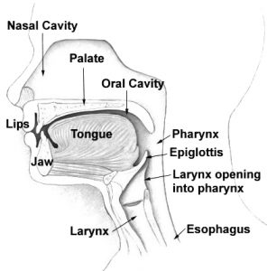
The pharynx is a component of the digestive system and respiratory system. It is a soft part above throat that links the nose and mouth to the esophagus, which is the tube that carries the food to the stomach. As part of the digestive systems, its muscular walls support in swallowing process and carrying food to the esophagus. Being part of the respiratory system, it enables movement of air from mouth and nose to the larynx in function of breathing.
Characteristics of the Pharynx
- It is a structure found in both vertebrates and invertebrates.
- The pharynx is approximately 5 inches in length.
- It is common to both the gastrointestinal and respiratory tracts and carries out functions for both systems.
Pharynx Location and Origin
The pharynx is essentially part of the throat and is located behind the nasal cavity and mouth and above the larynx and esophagus. It connects to the middle ear cavity on either side through the Eustachian tubes.
Pharynx Structure
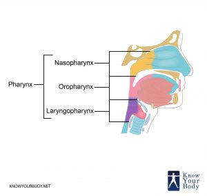
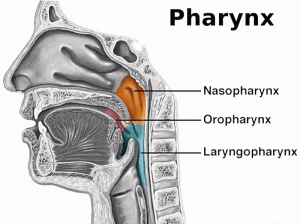
The structure of pharynx is divided into three components – Nasopharynx, Oropharynx, and Laryngopharynx.
Nasopharynx
The nasopharynx is the upper part of the pharynx that stars from the skull base to the upper layer of the soft palate. It comprises space between the soft palate and internal nares and is above the oral cavity. It consists of adenoids, which are also called as pharyngeal tonsils that are lymphoid tissue structure situation in nasopharynx’s posterior wall.
The mucus or polyps can obstruct the nasopharynx and can lead to congestion because of infection in the upper respiratory system. It includes an auditory tube that connects the pharynx to the middle ear and opens at the pharyngeal opening of the auditory tube. The closing and opening of auditory tubes lead to equalization of barometric pressure in the middle ear.
Oropharynx
This part of pharynx rests behind oral cavity, starting from uvula to hyoid bone. It opens anteriorly from the Isthmus faucium into the mouth, and its lateral wall rests between the palatopharyngeal arch and palatoglossal arch, which is the palatine tonsil. Its anterior wall comprises the tongue base, epiglottic vallecula, and the lateral wall is prepared of tonsillar fossa, tonsil, and tonsillar pillars. Its superior wall comprises interior surface from the soft palate and the uvula. It enables passing of air and food through a layer of connective tissue known as epiglottis.
Laryngopharynx
Also called as hypopharynx, it is the caudal component of the pharynx. It links to the esophagus and rests in the inferior to the epiglottis. It diverges into digestive and respiratory pathways. From this point, it is continuous from the esophagus posteriorly. It carries the fluids and foods to the stomach and air enters into the larynx anteriorly. The three major parts of laryngopharynx are – the pyriform sinus, posterior pharyngeal wall, and postcricoid area.
Pharynx Anatomy and Muscles
The pharynx is a tube made of muscles. There are two kinds of muscles that create the pharynx walls – circular and longitudinal. Both forms of muscles are innervated from the vagus nerve, excluding the stylopharyngeus that is innervated from the glossopharyngeal nerve.
Circular Muscle
The circular muscle contracts from the inferior to superior to obstruct the lumen and boost the bolus of food inferiority into the esophagus. These muscles are loaded with glasses and are an incomplete muscular circle, linking to the neck structures. The various types of circular muscles are:
- Superior Pharyngeal Constrictor – This muscle is present in the oropharynx. It extends from the mandible, pterygomandibular raphe, pterygoid hamulus, and from the side part of the tongue to the pharyngeal tubercle.
- Middle Pharyngeal Constrictor – It is present in laryngopharynx. It extends from stylohyoid ligament and hyoid horns to pharyngeal raphe.
- Inferior Pharyngeal Constrictor – It is identified in the laryngopharynx and has two parts. The inferior component also known as cricopharyngeus includes horizontal fibers that are linked to cricoid cartilage. The superior component also known as thyropharyngeus includes oblique fibers that link to cricoid cartilage.
Longitudinal Muscle
The pharynx includes longitudinal muscles that widen and shorten pharynx and boost the larynx while swallowing. The three forms of longitudinal muscles are:
- Palatopharyngeus – This is the hard palate of the oral cavity and innervated from the vagus nerve.
- Stylopharyngeus – It begins with the styloid process belonging to the temporal bone of the pharynx. It innervates from the glossopharyngeal nerve.
- Salpingopharyngeus – It starts from the Eustachian tube and moves to the pharynx. The innervation is obtained from the vagus nerve. Besides leading to swallowing, it opens the Eustachian tube to balance the pressure of the atmosphere and middle ear.
Pharynx Functions
The main functions of the pharynx are respiratory and digestive in nature.
- The muscles of the pharynx help in the mechanism of swallowing and provide a pathway for food to move from the mouth to the esophagus. When the tongue pushes the masticated food to the back of the mouth, the windpipe closes and the food is moved into the pharynx. The muscles work to swallow the food and move it into the esophagus.
- The pharynx allows the passage of inhaled air from the nose into the larynx, the windpipe and then the lungs. The isthmus which is the pathway that connects the oral and nasal pharynxes, allows humans to breathe through both the nose and the mouth.
- The pharynx works in tandem with other muscles related to speech to produce sounds and also plays a role in sound resonation.
- As it is connected to the ear cavity on either side through the Eustachian tubes, this helps in equalizing the pressure created by air on the eardrums.
- The lymphoid tissues found in the pharynx play a small role in regulating the immune system of the body. The lymphoid tissues that make up the tonsils monitor the particles and microbes that enter the nose or throat and prevents them from mixing with the air that goes into the lungs or the food that goes into the stomach.
Pharynx Innervation
The pharynx is largely innervated by the branches of the vagus nerve, the glossopharyngeal nerve and the fibers of the superior cervical ganglion. The stylopharyngeus is the only muscle to be innervated by the glossopharyngeal nerve (CN IX). The other muscles of the pharynx (both circular and longitudinal) are innervated by the vagus nerve (CN X).
Pharynx Blood Supply
The branches of the external carotid artery form the arterial blood supply. The pharyngeal venous plexus is the vein that drains the blood into the internal jugular.
Pharynx Clinical Complications
- Pharyngitis – This refers to the inflammation of the pharynx which causes swelling, redness, and pain during swallowing. Severe cases require antibiotics prescribed by the doctor.
- Pharyngeal Cancer – This form of throat cancer creates bleeding, pain in the neck and throat, can have lumps which make it difficult to swallow and can even result in deafness.
- Laryngopharyngeal reflux – This is common for those suffering from gastric problems when the stomach acids rise up via the esophagus.
- Tonsillitis – The palatine tonsils sometimes get inflamed with a bacterial or viral infection and then get really uncomfortable or painful. The lymphatic nodes become enlarged and there could be a difficulty in speaking or swallowing. A tonsillectomy is usually needed here.
- Throat paralyzes – This situation can occur because of an infection in the throat and due to diseases like rabies, polio, and diphtheria.
- Pharynx Cancer – This is another clinical complication of the pharynx that is caused because of lumpy feeling in the neck or throat and can lead to nasal obstruction, throat pain, ear pain, bleeding, problems swallowing, and sometimes deafness.
Pharyngitis Treatment
The most common disease caused in Pharynx is pharyngitis. If you are experiencing such problem then your doctor will examine the throat for having any gray or white patches. The doctor would even check the nose and ear for swollen lymph nodes near the side of the neck. If the doctor identifies any problem then he will perform the throat culture.
After the throat culture, the doctor would also perform a blood test to identify the actual cause of pharyngitis. The blood sample is sent for testing to determine that whether you are suffering from mononucleosis or suffering from any other type of infection.
Most throat problems get better within a week. Usually, antibiotics are prescribed in order to confirm that the risk of the ailment is completely rectified. Besides the painkillers, the doctor suggests to:
- Consume cool and soft food having more liquid consistency.
- Avoid smoking
- Such hard sweets, ice lollies, ice cubes, and lozenges
- Gargle on a result basis with lukewarm and salty water
Pharynx Pictures
Frequently Asked Questions
What exactly are the tonsils?
The tonsils are structures made of lymphoid tissue that are located in the pharynx. It has an immune-related function which mainly monitors the air, fluids, and food entering the nose and mouth.
What are the different types of tonsils in the human body?
The pharyngeal tonsils or adenoids are located in the nasopharynx. The tubal tonsil is located in the submucosa layer near the opening of the Eustachian tube. The palatine tonsils are on either side of the oropharynx in between the palatine arches. The lingual tonsils are all the way at the back of the tongue.
What is Waldeyer’s Ring?
This is also known as the pharyngeal lymphatic ring. Waldeyer’s Ring is a collective term for lymphoid tissue which includes the four tonsils named above. It is basically an uninterrupted ring that has protective functions in nature in keeping out microbes and irritants that are inhaled or ingested.
What is a tonsillectomy?
Tonsillitis refers to inflammation of the palatine tonsils. The tonsillectomy is a surgical procedure that removes the tonsils when they become inflamed and uncomfortable.
Are there risks associated with a tonsillectomy?
The tonsillectomy is a very common procedure. However, there could be a few associated risks such as swelling, infections, bleeding and extreme reactions to the anesthetics used.
What happens to the adenoids as you age?
The adenoids are at their largest during childhood, roughly between the ages of 3-5. After that, it regresses in size.
The pharynx is a tubular structure that originates at the back of the mouth and nose and extends till the larynx and esophagus. It has both respiratory and digestive functions by providing a pathway for filtered air to reach the lungs and food to reach the stomach through the esophagus.


No comments yet.