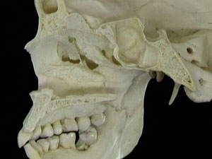
Frontonasal duct Definition It is the passage that moves down from the frontal sinus and opens into the ethmoidal infundibulum. The frontal sinus drains into the middle meatus through the frontonasal duct. Each of the frontal sinuses consists of this passage. Frontonasal duct Location This duct is located in a narrow vertical position. It may […]
Last updated on February 1st, 2018 at 12:43 pm
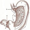
Angle of His Definition It is the acute angle that is formed between the entryway to the stomach and the esophagus, or the passage between the pharynx and the stomach. It is also known as the “His angle”. Angle of His Anatomy The angle is formed by the fibers of collar sling and the circular […]
Last updated on March 16th, 2018 at 4:32 am
Renal lobe Definition It is a part of the kidney that comprises of a renal pyramid as well as a renal cortex on top of it. The structure can be viewed in humans without a microscope. It is difficult to find it out in animals without the aid of such an apparatus. This is due […]
Last updated on April 11th, 2018 at 6:45 am
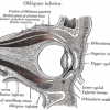
Superior tarsal Definition It is the smooth muscle lying next to the levator palpebrae superioris muscle whist assists in raising the upper eyelid. It is also known as “Muller muscle”, a term that is also used to refer to a part of the ciliary muscle. Superior tarsal Location This well-defined muscle starts from the bottom […]
Last updated on November 14th, 2018 at 8:08 pm
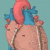
Anterior interventricular sulcus Definition It refers to a groove or furrow on the anteriosuperior cardiac surface, which designates the position of the septum between the right and left ventricles. It is one of the two grooves demarcating the two ventricles of the heart, the other being the posterior interventricular sulcus. Anterior interventricular sulcus Synonyms This […]
Last updated on November 14th, 2018 at 8:15 pm
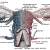
Cardinal Ligament Definition It is a major uterine ligament of the human body. The female body contains a pair of these structures. It is part of a visceral pelvic fascial thickening. Cardinal Ligament Synonyms It is also known by many different names like Lateral Cervical Ligament Transverse Cervical Ligament Mackenrodt’s Ligament Cardinal Ligament Location The […]
Last updated on August 19th, 2018 at 6:31 am
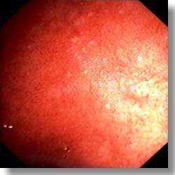
Duodenal bulb Definition It is the part of the duodenum that is closest to the stomach. Duodenal bulb Appearance This duodenal structure is about 5 cms long. It is round in shape and has a smoother surface than the other parts of the duodenum that comprise of villi (tiny hair-like mucosal projections), as well as […]
Last updated on September 2nd, 2018 at 8:38 am
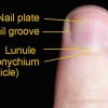
Eponychium Definition It refers to a tiny epithelium band that stretches onto the nail base from the posterior section of the nail. It is actually the terminal point of the proximal fold that rolls back to cast off an epidermal skin layer over a nail plate that has been formed freshly. Eponychium Synonyms It is […]
Last updated on July 28th, 2018 at 8:12 am
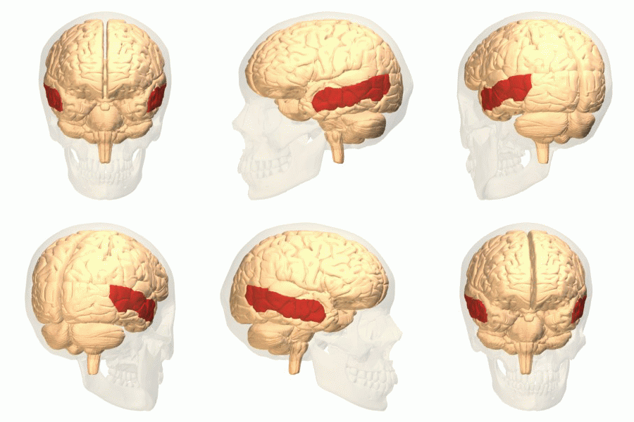
Middle temporal gyrus Definition It refers to a gyrus, meaning an elevation or a convex fold, on the temporal lobe in the surface of the brain. This structure should not be confused with medial temporal lobe. It is abbreviated as MTG or even GTm. GTm refers to the fact that it is also often referred […]
Last updated on August 31st, 2017 at 11:55 am
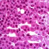
Zona reticularis Definition It is the innermost of the three tissue layers of the cortex in the adrenal gland. It comprises of cylindrical masses of epithelial cells that are arranged in an irregular, netlike fashion. The other two adrenal cortex layers, lying above this layer, are Zona glomerulosa and Zona fasciculata. Zona reticularis Location It […]
Last updated on August 31st, 2017 at 12:06 pm


